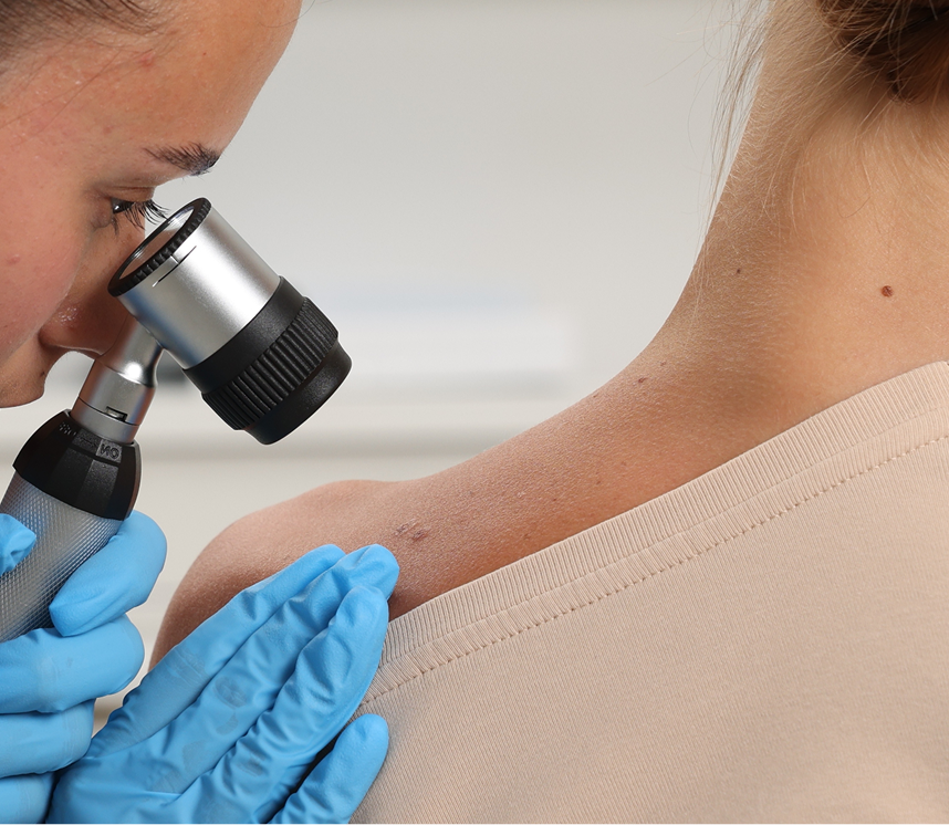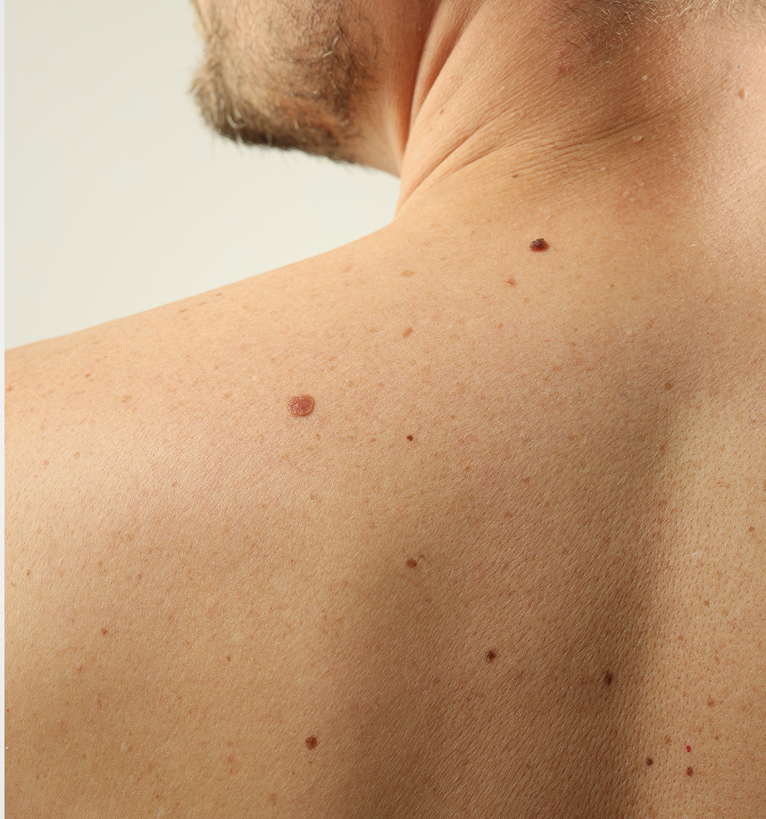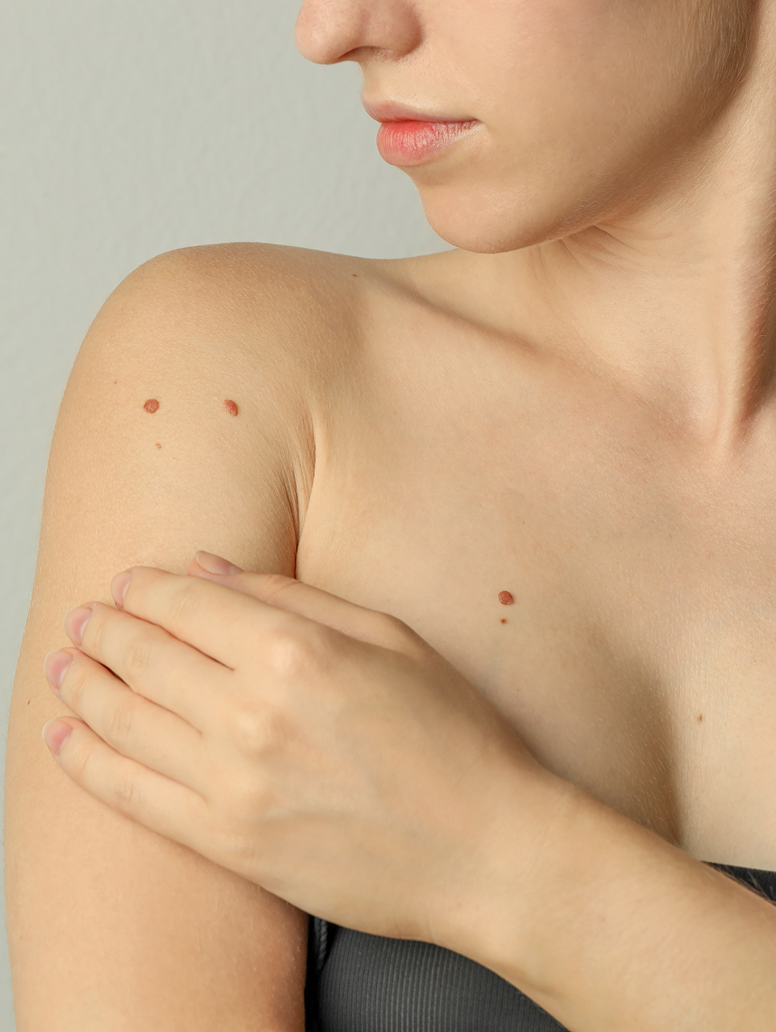
Changes in your skin can be a significant source of concern. Our expert team is dedicated to providing thorough diagnostic services, including precise skin biopsies, to accurately identify and address a wide range of dermatological conditions. With advanced techniques and a commitment to patient comfort, we ensure you receive the clearest answers and the most appropriate care for your skin health.
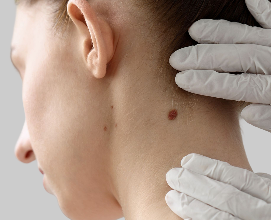

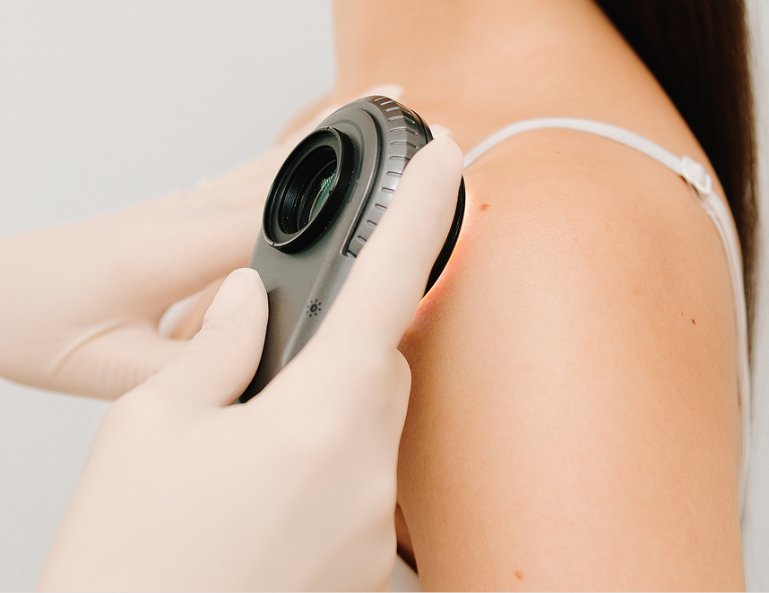
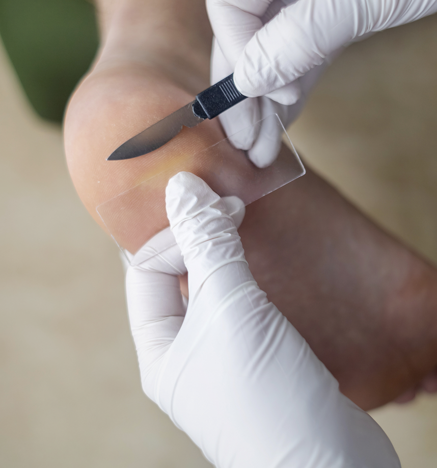
Shave Biopsy: This technique involves using a small razor blade to “shave” off the outermost layers of skin, including the epidermis and a portion of the dermis. It’s commonly used for lesions that are raised above the skin surface, such as skin tags, seborrheic keratoses, and certain types of basal cell carcinoma.
Punch Biopsy: For conditions requiring a deeper tissue sample, a punch biopsy is performed. A circular tool, similar to a small cookie cutter, is rotated into the skin to remove a cylindrical core of tissue, including the epidermis, dermis, and sometimes a portion of the subcutaneous fat. This method is often used for suspicious moles, inflammatory rashes, and when assessing the depth of a lesion.
Excisional Biopsy: This method involves surgically removing the entire lesion, along with a small margin of surrounding healthy skin. It is typically performed for lesions in which complete removal is desired, either for definitive diagnosis or as treatment for conditions such as melanoma, ensuring that all abnormal cells are removed. The wound is then closed with sutures.
Incisional Biopsy: In cases where a lesion is large or located in a cosmetically sensitive area, an incisional biopsy may be performed. This technique involves removing only a representative portion of the lesion, allowing for diagnosis without immediately removing the entire growth. If the biopsy reveals malignancy, further treatment or a subsequent excisional procedure may be necessary.